
Figure 5.
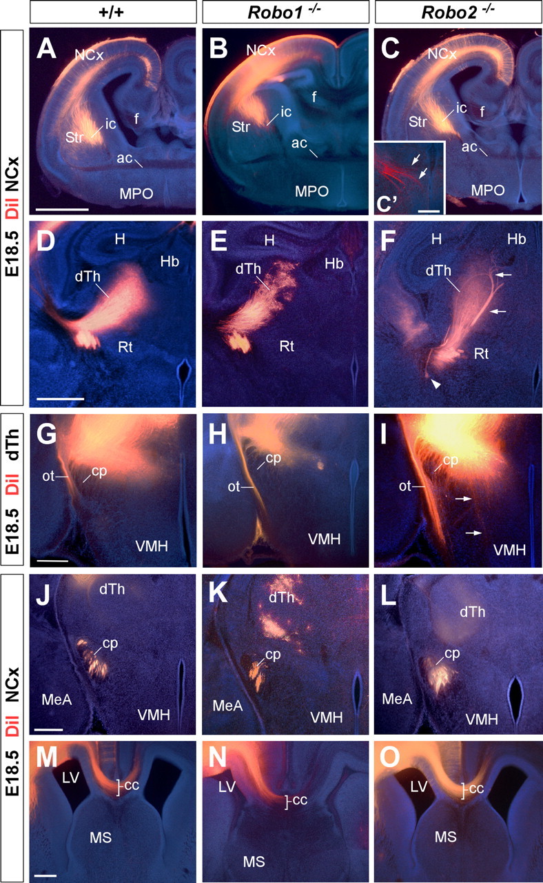
Axon guidance defects in the forebrain of Robo2 single-mutant mice. Coronal sections through the telencephalon of E18.5 brains with DiI implanted in the neocortex (NCx) (A–F, J–O) or in the dorsal thalamus (dTh) (G–I) of wild-type (A, D, G, J, M), and Robo1 (B, E, H, K, N) and Robo2 (C, F, I, L, O) mutant mice, showing computer-generated overlays of DiI-labeled axons and Hoechst counterstain. A–C′, Coronal sections showing labeled DiI axons extending from the cortex into the internal capsule (ic) in wild-type (A), and Robo1 (B) and Robo2 (C, C′) mutant mice. D–F, Abnormal defasciculation (arrowhead) and targeting (arrows) of corticothalamic axons at the dorsal thalamus of Robo2 mutant mice (F). In Robo1 mutant mice, labeled axons extend from the cortex into the dorsal thalamus normally. G–I, Coronal sections through the caudal diencephalon showing dorsal thalamic fibers abnormally entering the hypothalamus in Robo2 mutant mice (I). No guidance defects were observed in the thalamocortical axons in Robo1 mutant mice (H) compared with wild type (G). J–L, Coronal sections showing corticospinal labeled axons at the cerebral peduncle (cp) of wild-type (J), and Robo1 (K) and Robo2 (L) mutant mice. Note the abnormal ventral position of the cerebral peduncle and the defasciculation of axons in Robo2 mutant mice. M–O, Rostral coronal sections showing corticocortical axons at the corpus callosum (cc) of wild-type (M), and Robo1 (N) and Robo2 (O) mutant mice. No guidance defects were observed in either of the Robo single mutants. ac, Anterior commissure; Str, striatum; f, fimbria; MPO, medial preoptic area; H, hippocampus; Rt, reticular thalamic nucleus; Hb, habenula; VMH, ventromedial hypothalamic nucleus; MeA, medial amygdala nucleus; MS, medial septum; ot, optic tract; LV, lateral ventricle. Scale bars: A–C, 1 mm; C′, 200 μm; D–F, 500 μm; G–O, 300 μm.