
Figure 9.
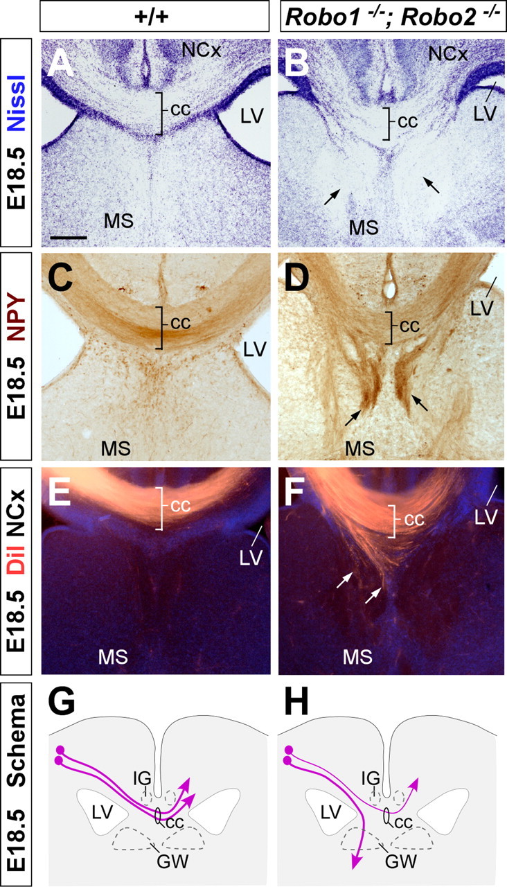
Abnormal development of the corpus callosum in Robo1;Robo2 double-mutant mice. Coronal sections through the telencephalon of E18.5 fetuses showing Nissl stain (A, B) and NPY (C, D) immunohistochemistry in wild-type (A, C) and Robo1;Robo2 double-mutant mice (B, D) mutant mice. A–D, Nissl staining and NPY immunohistochemistry demonstrates that Robo1;Robo2 double-mutant mice have a small corpus callosum (cc) and that large ectopic bundles of axons form on either side of it (B, D, arrows). E, F, Coronal sections through the telencephalon of E18.5 fetuses with DiI implanted in the neocortex (NCx), showing DiI-labeled corticocortical axons extending through the corpus callosum in wild-type (E) and Robo1;Robo2 double-mutant (F) mice. Note that many axons are abnormally directed ventrally before they reach the midline (F, arrows). G, H, The schemas summarize the pathways followed by corticocortical axons through the corpus callosum in wild-type (G) and Robo1;Robo2 double-mutant (H) mice. MS, Medial septum; LV, lateral ventricle; GW, glial wedge; IG, indusium griseum. Scale bars: A–F, 200 μm.