
Figure 8.
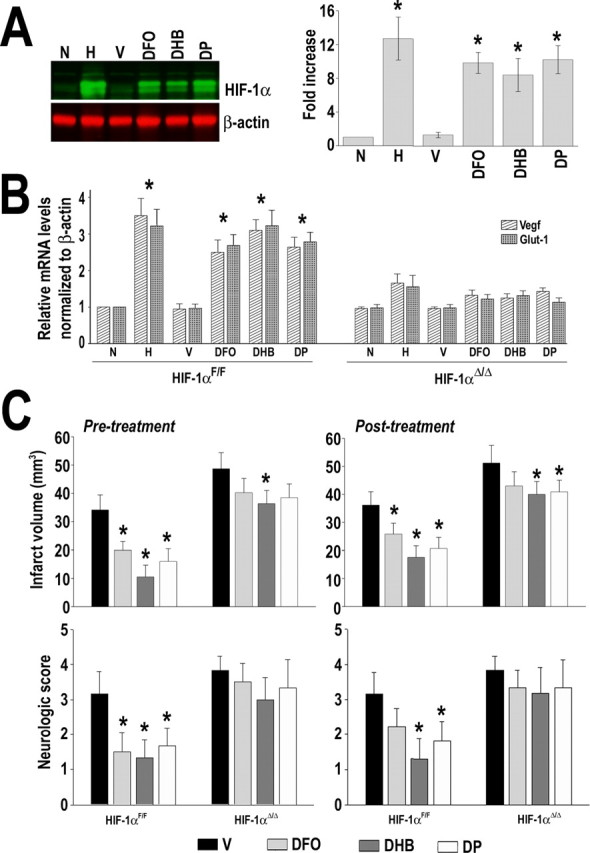
Effect of prolyl hydroxylases inhibitors on ischemic brain damage. A, Western blot analysis of HIF-1α in brain cortex of animals treated with vehicle intraperitoneally (V), DP (20 mg/kg, i.p.), DHB (200 mg/kg, i.p.), and DFO (300 mg/kg, s.c.). Animals were killed 6 h after drug administration. Normoxic (N) and hypoxic (H) (8% O2 for 4 h) animals were used for comparison. B, RT-PCR analysis of Vegf and Glut-1 in brain cortex at 24 h after treatment of animals (HIF-1αF/F and R1-HIF-1αΔ/Δ) as indicated in A. C, Pretreatment of HIF-1αF/F and R1-HIF-1αΔ/Δ mice with the indicated drugs was performed 6 h before the ischemic insult, and posttreatment began 6 h after MCAo. Evaluation of infarct volume and neurological deficit was made 4 d after the onset of ischemia. Data are presented as mean ± SD (n = 6). *p ≤ 0.05 compared with normoxia (N) or vehicle (V).