Figure 1.
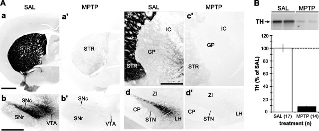
Light microscopic immunoperoxidase and Western immunoblot analyses of TH in the striatum, GP, and STN of MPTP-treated mice. A, Representative light micrographs of DAT-immunostained basal ganglia tissue sections from SAL- and MPTP-treated mice. In STR (a, a′), GP (c, c′), and STN (d, d′), MPTP treatment resulted in the loss of DAT-labeled dopaminergic fibers. In SN (b, b′), the same MPTP treatment caused a massive loss of DAT-positive dopaminergic neurons and neuropil. Scale bars: Aa (for Aa, Aa′), Ab (for Ab, Ab′), Ac (for Ac, Ac′, Ad, Ad′), 500 μm. CP, Cerebral peduncle; IC, internal capsule; LH, lateral hypothalamus; SNc, substantia nigra pars compacta; SNr, substantia nigra pars reticulata; VTA, ventral tegmental area; ZI, zona incerta. B, Top, A representative immunoblot of the striatal tissue samples from two SAL-treated and two MPTP-treated mice, demonstrating a profound decrease in the expression level of TH, a marker for dopaminergic terminals. Bottom, Quantification of the intensity of TH-immunoreactive bands from SAL- (n = 17) and MPTP-treated (n = 14) mice, expressed as mean ± SEM. Note that the MPTP treatment causes >90% reduction in TH level on average.

