
Figure 1.
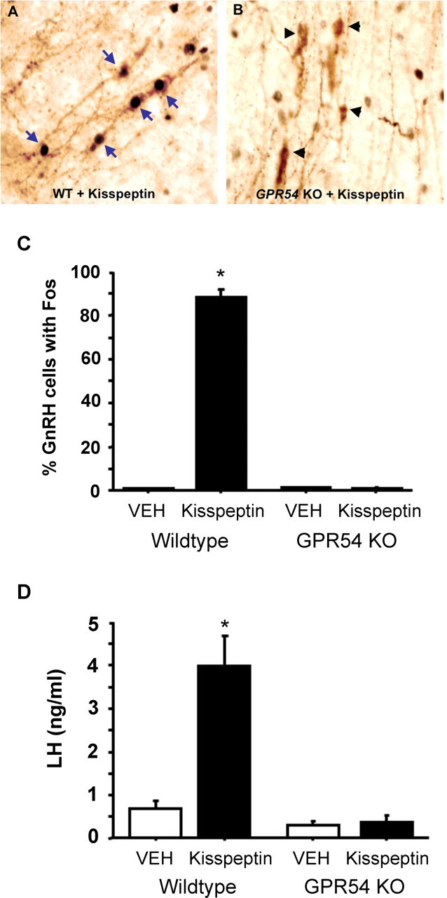
Representative photomicrographs of GnRH-immunoreactive neurons in the forebrain of gonad-intact WT (A) and GPR54 KO (B) male mice infused intracerebroventricularly with 1 nmol of kisspeptin. Fos immunoreactivity (labeled black) is visible in the nucleus of GnRH cells (labeled brown) of WT but not GPR54 KO mice. Blue arrows denote example GnRH neurons colabeled with Fos; black arrowheads denote GnRH neurons lacking Fos. C, Mean (±SEM) percentage of GnRH neurons colabeled with Fos in WT and GPR54 KO males treated with kisspeptin or vehicle (VEH). *p < 0.05, significantly different from vehicle-treated WT mice. D, Mean (±SEM) concentration of plasma LH in WT and GPR54 KO males 30 min after treatment with kisspeptin or vehicle (VEH). *p < 0.05, significantly different from vehicle-treated WT mice.