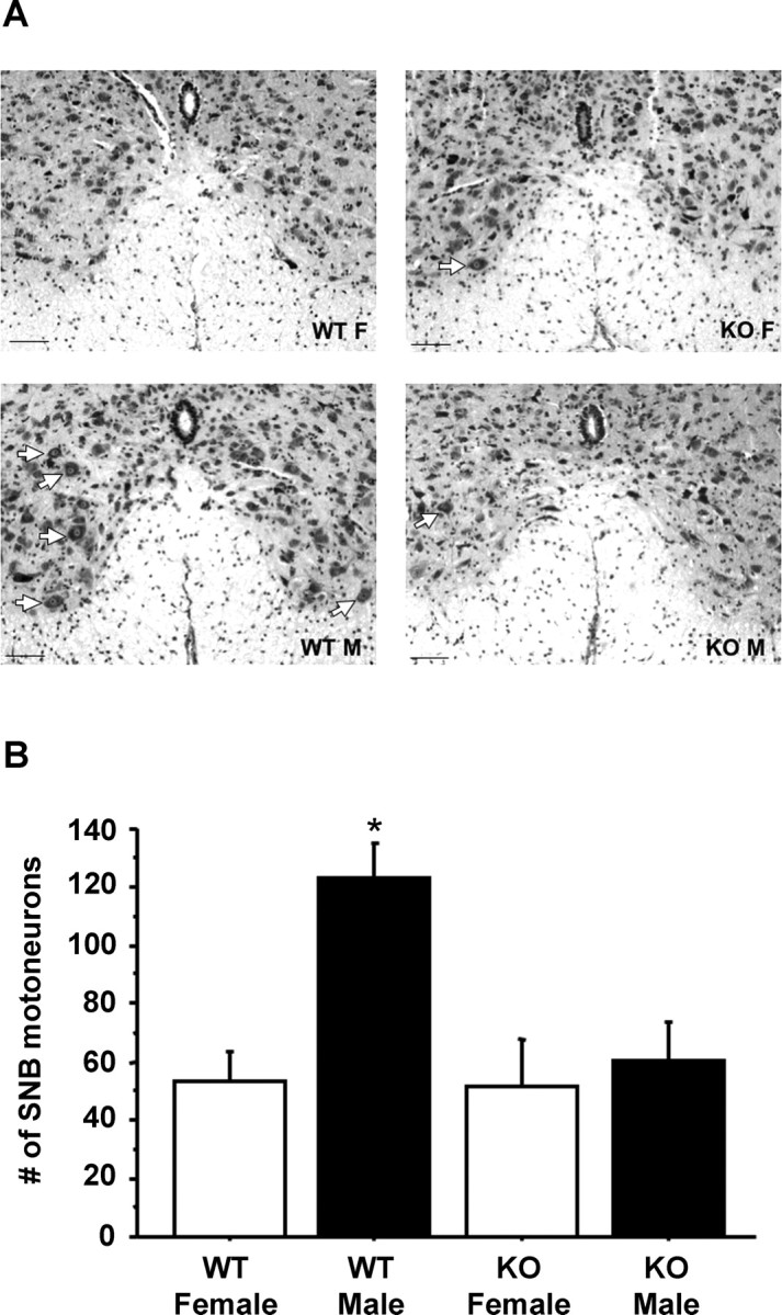
Figure 6.

A, Photomicrographs of the ventral horn of the spinal cord of representative male (M) and female (F) WT and GPR54 KO mice. Photomicrographs were taken at 20× magnification. White arrows denote example SNB motoneurons, noticeable primarily in the WT males. Scale bar, 50 μm. B, Mean (±SEM) SNB motoneuron counts in adult WT and GPR54 KO mice of both sexes. *p < 0.01, significantly different from all other groups.
