
Figure 2.
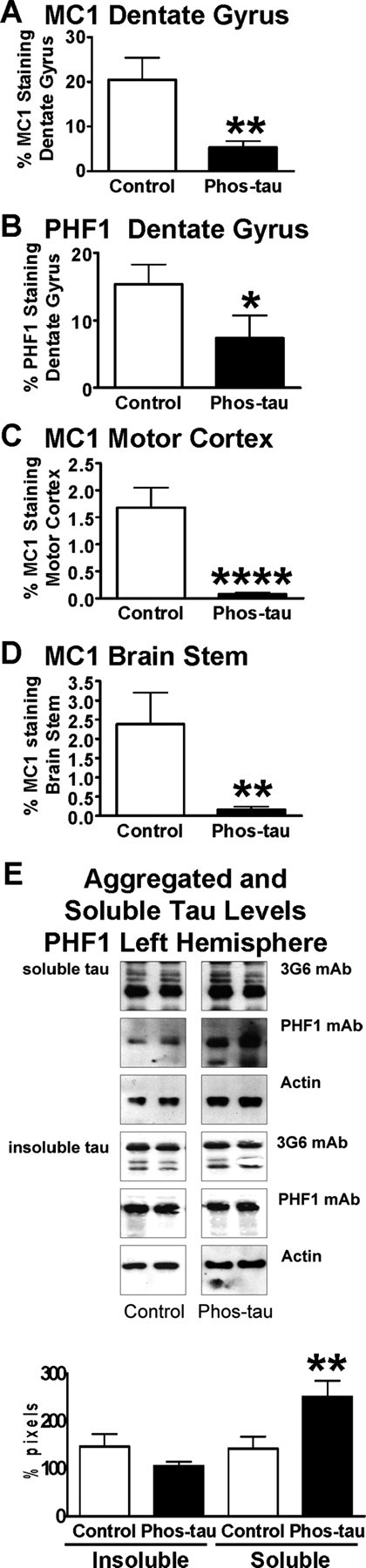
The vaccine reduces tau aggregates in the brains of P301L tangle mice at 5 months of age. A, Quantitative analysis of MC1 immunoreactivity within the granular layer of the dentate gyrus revealed a 74% reduction (**p < 0.01) in immunized mice compared with control Tg mice that received adjuvant alone. B, Likewise, PHF1 immunoreactivity within the granular layer of the dentate gyrus was reduced by 52% (*p < 0.05) in immunized mice compared with controls. C, Additional confirmation of a therapeutic effect was obtained by analysis of the motor cortex, in which MC1 neuronal staining was reduced by 96% (****p < 0.0001) compared with control Tg mice. D, Likewise, in the brainstem, MC1 neuronal staining was reduced by 93% (**p = 0.01) compared with control Tg mice. E, Densitometric analysis of PHF1 blots revealed a strong trend for reduction in insoluble tau (28% reduction; p = 0.09) and a significant increase in soluble tau (77% increase; **p = 0.01) in the immunized mice compared with control Tg mice, relative to total tau levels. Additional analysis of the ratio of soluble tau to insoluble tau indicated a significant increase in the immunized group on PHF1 blots (89% increase; p = 0.01), suggesting a mobilization of tau from its insoluble form to soluble form in these treated animals. The panel shows representative blots from control and Phos-tau-immunized mice. The PHF1 antibody recognizes phosphorylated serines 396 and 404 located outside the microtubule-binding repeat on the C terminus of PHF tau protein. Antibodies against total tau (3G6) and actin were used as controls. The same amount of protein was loaded in each line. Mean values are presented with SEM.