
Figure 5.
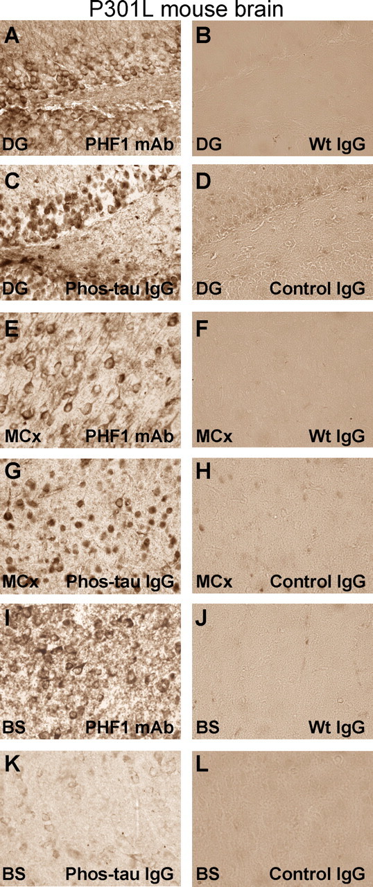
Purified antibodies from immunized mice stain tau aggregates/tangles in neuronal cell bodies in P301L mice. A–L, Adjacent coronal brain sections through the dentate gyrus (A–D), motor cortex (E–H), and brainstem (I–L; immediately below the Aqueduct of Sylvius) in a P301L transgenic mouse with tau pathology. A, The PHF1 antibody reveals the typical staining of tau aggregates/tangles in neuronal cell bodies as previously reported in this model (Lewis et al., 2000). Most of the staining is associated with the neuronal plasma membrane. B, D, Pooled mouse IgG from wild-type mice [Wt IgG (Sigma)] (B) or antibodies from mice immunized with the adjuvant only (Control IgG) (D) do not decorate neurons in the dentate gyrus. C, Antibodies from mice immunized with the Phos-tau peptide, which contains the PHF1 epitope, lack extensive dendritic staining, but stain primarily neuronal cell bodies within the dentate gyrus, but the pattern is not identical to the PHF1 staining. E, G, I, K, Similar staining pattern as in A and C is obsered in the motor cortex (E, G) and brainstem (I, K) after immunoreactivity with the PHF1 antibody and the polyclonal antibody from an immunized mouse, respectively. However, this particular polyclonal antibody stained neurons in the brainstem less intensely than in the dentate gyrus and motor cortex. F, H, J, L, These images depict adjacent coronal sections to those shown in E, G, I, and K, which were stained with purified antibodies from Tg control mice that received adjuvant alone (Control IgG) or pooled mouse IgG (Wt IgG). Staining with those antibodies resulted in minimal or no staining. No immunostaining was observed in wild-type mice with the antibodies purified from immunized mice (data not shown). These findings indicate that the immunized mice generate antibodies that specifically recognize pathological tau aggregates in the P301L mouse. Staining was performed as detailed in Materials and Methods with PHF1 and purified IgG used at a 1:250 and 10 μg/ml dilutions, respectively. Original magnification, 400×.