Figure 6.

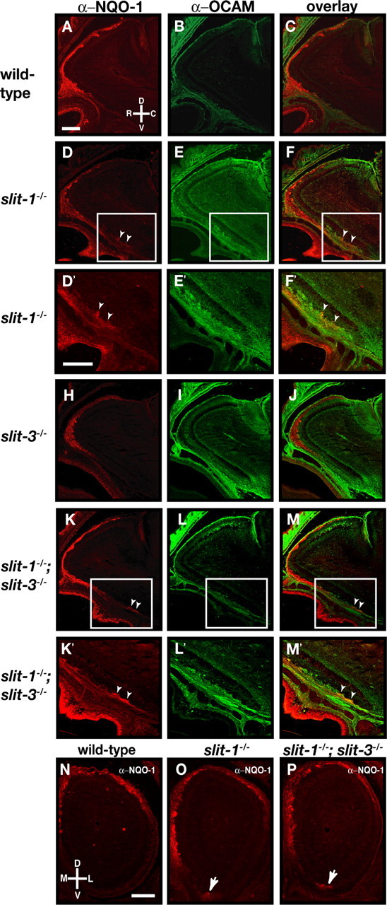
Loss of zonal targeting of NQO-1-expressing olfactory sensory neuron axons within the olfactory bulb in slit-1−/− mice. A–M, Parasagittal sections of olfactory bulbs from P0 wild-type (A–C), slit-1−/− (D–F), slit-3−/− (H–J), and slit-1−/−;slit-3−/− (K–M) mice stained with anti-NQO-1 (A, C, D, F, H, J, K, M) and anti-OCAM (B, C, E, F, I, J, L, M). In wild-type animals, NQO-1-expressing axons are restricted to the rostrodorsal region of the OB (A, C) and OCAM-expressing axons target to the ventral region of the olfactory bulb (B, C). Whereas NQO-1-expressing axons are properly targeted to the dorsal region of the olfactory bulb in slit-3−/− mice (H, J), a subset of NQO-1-expressing axons mistarget to the most ventral region of the olfactory bulb in slit-1−/− mice (D, F) (arrowheads). In slit-1−/−; slit-3−/− mice, NQO-1-expressing axons are also observed in the ventral region of the olfactory bulb (K, M) (arrowheads). High-powered magnifications of ectopically projecting NQO-1-expressing axons (insets in D, F, K, M) are shown in D′, F′, K′, and M′. n = 8 wild type, n = 5 slit-1−/−, n = 8 slit-3−/−, and n = 7 slit-1−/−; slit-3−/−. N–P, Coronal sections at similar rostrocaudal levels of olfactory bulbs isolated from P0 (N–P) wild-type (N), slit-1−/− (O), and slit-1−/−; slit-3−/− (P) mice stained with anti-NQO-1 (N–P). In wild-type mice, NQO-1-expressing axons innervate the dorsomedial region of the olfactory bulb. In slit-1−/− and slit-1−/−; slit-3−/− mice, a subset of NQO-1-expressing axons are mistargeted to the ventral region of the olfactory bulb (arrows). n = 8 wild type, n = 8 slit-1−/−, and n = 5 slit-1−/−; slit-3−/−. D, Dorsal; V, ventral; L, lateral; M, medial; R, rostral; C, caudal. Scale bars, 250 μm.
