
Figure 1.
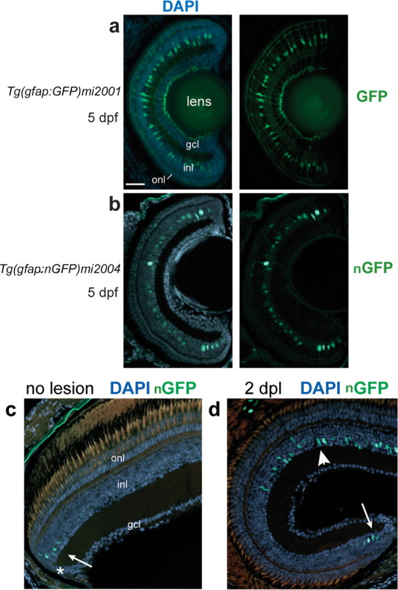
GFP+ Müller glia in Tg(gfap:GFP)mi2001 and Tg(gfap:nGFP)mi2004 transgenic zebrafish. a–d, Retinal cryosections from larval (a, b) or adult (c, d) transgenic zebrafish counterstained with DAPI (blue). a, In the Tg(gfap:GFP)mi2001 and mi2002 cytoplasmic GFP lines, Müller glia cell bodies in the inner nuclear layer (inl) extend radial processes toward the outer nuclear layer (onl) and ganglion cell layer (gcl). b, In the Tg(gfap:nGFP)mi2004 line, reporter expression is restricted to nuclei of Müller glia in this young larval fish, at 5 dpf. The GFP expression in this preparation is visualized by immunofluorescence with an anti-GFP antibody. c, In adult Tg(gfap:nGFP)mi2004 fish, endogenous nuclear-targeted GFP expression is detected only in immature Müller glia nuclei (arrow) near the CMZ (*). d, In Tg(gfap:nGFP)mi2004 fish at 2 d after destruction of photoreceptors by exposure to high-intensity light (days postlesion), endogenous nuclear-targeted GFP expression is upregulated in Müller glia within the lesioned area (arrowhead) similar to immature Müller glia in newly generated retina at the ciliary margin (arrow). Scale bar: (in a) a, b, 40 μm; c, d, 30 μm.