
Figure 2.
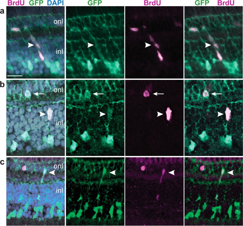
GFP+ Müller glia proliferate in the differentiated, growing retina. a–c, Retinal cryosections from juvenile Tg(gfap:GFP)mi2001 zebrafish labeled with BrdU (magenta) and the nuclear stain DAPI (blue). Representative images from two independent experiments, six fish per experiment, 7 d continuous exposure to BrdU. a, A radial chain of GFP+, BrdU+ cells (arrowhead) extends through the inner nuclear layer (inl) into the outer nuclear layer (onl). b, A single GFP+, BrdU+ cell in the INL (arrowhead) near a GFP+, BrdU+ cell in the ONL (arrow). c, Another example of a chain of GFP+, BrdU+ cells that spans INL and ONL (arrowhead). Scale bar: (in a) a, b, 15 μm; c, 20 μm.