
Figure 3.
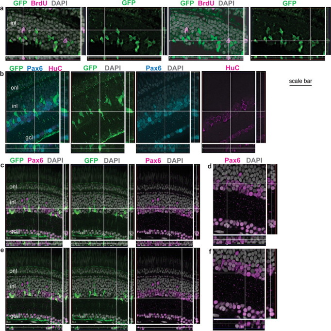
GFP+ Müller glia are colabeled with BrdU and Pax6 but not HuC. Retinal cryosections from juvenile Tg(gfap:GFP)mi2001 (a) and mi2002 (b–e) transgenic zebrafish. Reconstruction of optical slices (Z-stack) reconstructed and displayed in “cut view” through the thickness of the section in orthogonal planes. Sections were counterstained with DAPI (gray). a, This fish was exposed to BrdU for 7 d, and both of the BrdU+ nuclei (magenta) in the inner nuclear layer (inl) of this image are weakly GFP+ (green). b, This GFP+ cell (at the intersection of the crosshairs) is weakly immunoreactive for Pax6 (cyan) but not the neuronal marker HuC (magenta). As expected, many of the Pax6+, GFP− neurons (amacrine cells in the INL) and retinal ganglion cells in the ganglion cell layer (gcl) are double labeled with HuC. c–f, The crosshairs show two GFP+ (green) Müller glia with characteristic polygonal nuclei that express Pax6, shown at higher magnification in d and f, respectively. Scale bar: a, b, 30 μm; c, e, 40 μm; d, f, 27 μm. onl, Outer nuclear layer.