Figure 9.
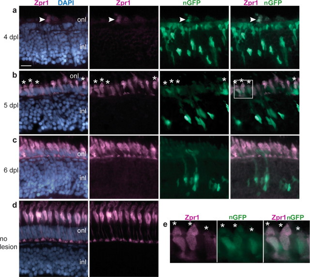
Regenerated cone photoreceptors are GFP+. a–d, Cryosections of adult zebrafish retinas labeled with zpr1 (magenta), GFP (green), and DAPI (blue). a–c, Light-lesioned retinas from Tg(gfap:nGFP)mi2004 fish. a, At 4 dpl, a few cells with weak zpr1 expression are in the outer nuclear layer (onl) in the lesioned area. A GFP+ cell is colabeled with zpr1+ (arrowhead). b, By 5 dpl, many zpr1+ immature regenerating cones are present in the ONL, and several are GFP+ (*). c, At 6 dpl, zpr1+ regenerating cones are more differentiated and are no longer labeled with GFP. d, Differentiated double cones labeled with zpr1 in an unlesioned retina. e, Magnified view of the white-boxed area in b shows colocalization of zpr1 and GFP in immature, regenerating cones (*). Scale bar: (in a) a–d, 15 μm. inl, Inner nuclear layer.

