
Figure 1.
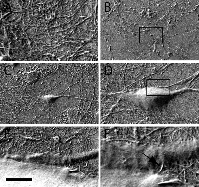
Scanning electron microscopy images of cultured hippocampal neurons on SWNTs. A, High-magnification micrograph showing SWNT details. B–D, Subsequent micrographs at higher magnifications of neurons grown on SWNTs (10 d). Same sample as in A is shown. Note the healthy morphology of neurons and the outgrowth of neurites attaching to the SWNT surface. E, F, Details of the framed area in D. At higher magnifications, the intimate contacts between bundles of SWNT and neuronal membrane are clearly shown. Scale bar (in E): A, 1 μm; B, 200 μm; C, 25 μm; D, 10 μm; E, 2 μm; F, 450 nm.