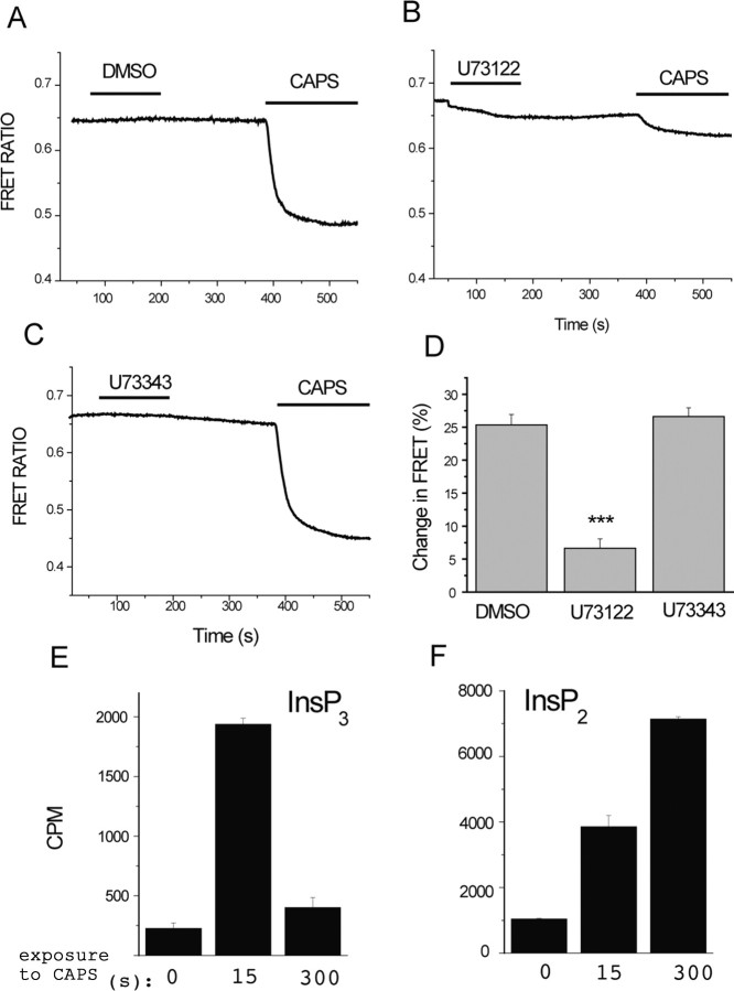
Figure 2.
Capsaicin activates PLC in TRPV1-expressing cells. A–D, Fluorescence was measured in HEK 293 cells expressing TRPV1 and the CFP- and YFP-tagged PLCδ1 PH domains, as described in Materials and Methods. Cells were pretreated with DMSO (A), 3 μm U73122 (B), or 3 μm U73343 (C) for 2 min, and then 1 μm capsaicin (CAPS) was applied, as indicated by the horizontal lines. The extracellular medium contained 1 mm Ca2+. The change in FRET ratio (D) was measured by dividing the value 2 min after the application of capsaicin with the FRET ratio value before the application of capsaicin to result Q, and then 100 − (100 × Q) was plotted (n = 14–17). ***p < 0.005. E, F, HEK cells expressing TRPV1 were incubated with 3H-inositol, as described in Materials and Methods, and stimulated with 2 μm capsaicin for the time periods indicated. Inositol phosphates were extracted and separated on HPLC, and the radioactivity was plotted for IP3 (InsP3; E) and IP2 (InsP2; F).

