
Figure 5.
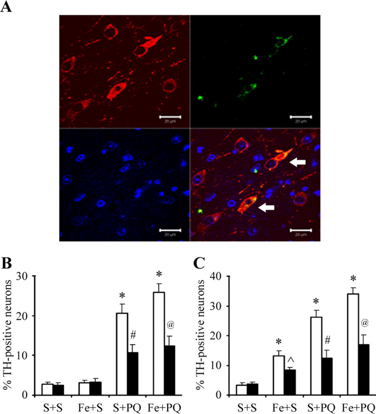
3-Nitrotyrosine immunopositive cells count in the SNpc. A, An example of localization (arrows) of 3-nitrotyrosine-immunopositive staining (green) within dopaminergic neurons (red). 4′,6-Diamidino-2-phenylindole (blue) was used to counterstain nuclei. Scale bars, 20 μm. B, C, Quantitative analysis of double labeling for TH with 3-nitrotyrosine in the SNpc of 2-month-old (B) and 12-month-old (C) mice. White bars, Vehicle; black bars, EUK-189 treated. Error bars indicate mean ± SEM. n = 3. *p < 0.001, significantly from saline plus saline plus vehicle group; p̂ < 0.05, significantly from neonatal iron fed plus saline plus vehicle group; #p < 0.001, significantly from saline plus paraquat plus vehicle group; @p < 0.001, significantly from neonatal iron fed plus paraquat plus vehicle group.