Figure 2.
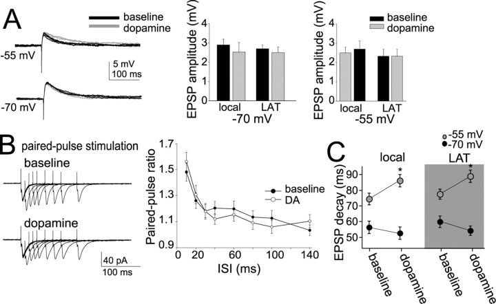
DA has minimal effects on EPSP amplitude. A, Application of DA (10 μm for 2–3 min) did not have a significant effect on the amplitude of a single LAT- or local-evoked EPSP, regardless of the membrane potential. Five traces in baseline and five after DA are overlayed and displayed for each panel. Black traces are baseline, and gray traces are after DA. B, Paired-pulse facilitation of EPSCs did not change after application of DA, examined over a range of frequencies. Displayed are overlays of averaged traces of paired stimulations at varying intervals. C, Application of DA resulted in a slower decay time of EPSPs (see A). This was observed specifically at depolarized membrane potentials. *p < 0.05, significant difference compared with baseline conditions (paired t test).

