
Figure 6.
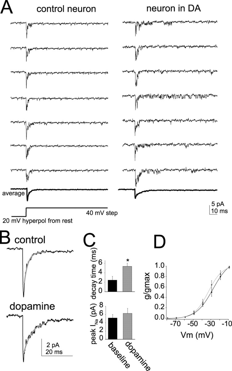
DA increases the activity of Na+ channels. A, Cell-attached recordings of multiple Na+ channels activated by a 40 mV step from 20 mV hyperpolarized to rest. Neurons exposed to DA (right) displayed a greater tendency for longer-lasting channel activity than nonexposed control neurons (left). B, The decay time of the averaged Na+ current was fit with a single exponential (gray). Neurons that were exposed to DA (bottom) displayed averaged current that decayed slower compared with control neurons. C, The decay time of the current was prolonged in neurons exposed to DA, and there was a trend toward increased average peak amplitude. The asterisk indicates a significant difference in a t test of control neurons compared with a separate group of neurons exposed to DA. D, Similar to nucleated patch recordings of INa, there was a shift in the activation of Na+ channels in neurons exposed to DA (gray).