
Figure 2.
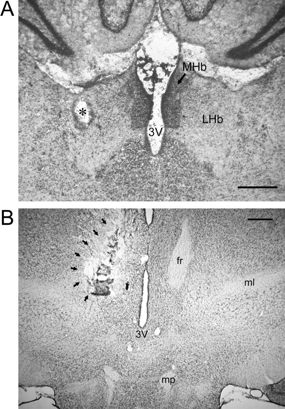
A, B, Photomicrographs illustrating placement of the stimulating electrode in the LHb (A) and tissue damage resulting from electrolytic lesion of the fasciculus retroflexus (B). The asterisk in A denotes the position of the tip of the stimulating electrode. The arrows in B outline the perimeter of a unilateral electrolytic lesion of the fasciculus retroflexus. MHb, Medial habenula; 3V, third ventricle; fr, fasciculus retroflexus; ml, medial leminscicus; mp, mammillary peduncle. Scale bars, 0.5 mm.