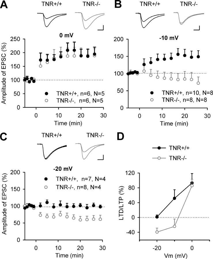
Figure 1.
Synaptic changes induced by pairing at 0, −10, and −20 mV in CA1 pyramidal cells of TNR deficient mice. A–C, Pairing of 1 Hz stimulation (applied at time 0) of Schaffer collateral/commissural fibers with membrane depolarization to 0 mV (A), −10 mV (B), or −20 mV (C) induces changes in EPSCs in TNR+/+ and TNR−/− mice. Data show means + SEM of normalized current amplitudes; n indicates the number of slices and N indicates the number of mice. Panels above LTP profiles show EPSCs recorded before and 30 min after pairing in TNR+/+ and TNR−/− mice. Calibrations: A, 10 ms, 100 pA; B, C, 10 ms, 50 pA. D, The levels of potentiation or depression (above or below the baseline level) as a function of membrane voltage (Vm) and genotype.

