Figure 3.

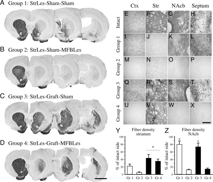
Ascending dopaminergic projections and fiber innervation provided by the graft. Four groups of animals were followed in this experiment. A, I–L, In group 1, the animals received a partial lesion depleting mainly the nigrostriatal projections to the dorsal and lateral striatum, leaving the mesocorticolimbic projections to the ventromedial striatum, nucleus accumbens, septum, and cortex intact. B, M–P, Group 2 consisted of animals that received a second lesion, which depleted the mesocorticolimbic pathway removing nearly all of the midbrain dopamine input to the forebrain areas. C, D, Q–X, Groups 3 (C, Q–T) and 4 (D, U–X) were animals that received transplants of fetal ventral mesencephalic tissue in multiple tracts to provide widespread reinnervation in the striatum in partially and completely denervated animals, respectively. In group 3, reinnervation of the striatal areas was sufficient to reinstitute the dopamine input systems to the forebrain; however, although the graft-derived fibers were equally abundant in the striatum in group 4, degeneration of the mesocorticolimbic fibers left the graft-derived dopamine input isolated (compare Q–T and U–X with E–H). Semiquantitative analysis of total TH-positive fiber density was measured in the striatum (Y) and the nucleus accumbens (Z). Ctx, Cortex; Str, striatum. Scale bars: (in D) A–D, 3 mm; (in X) E–X, 250 μm. *Different from nongrafted control groups; +different from the corresponding sham lesioned group. Error bars indicate SEM.
