Figure 6.
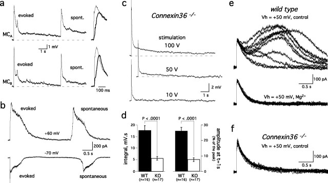
The intraglomerular network generates glutamatergic inputs onto mitral cell dendrites. a, Left, Spontaneous (spont.) depolarizations in a pair of mitral cells projecting to the same glomerulus had a similar time course to the evoked EPSPs. Note also that the depolarization were synchronous in the two cells. Right, Evoked events (thin traces) had faster rise times than spontaneous events (thick traces). b, In voltage clamp, spontaneous and ON-evoked currents also had similar shapes and reversed polarity at positive holding potentials (top trace). c, EPSPs were evoked by a single stimulation of the ON at different intensities in a mitral cell from a connexin36 knock-out mouse. d, Comparison of the integral (left) and decay (right) of EPSPs evoked in wild-type (WT) or Cx36−/− (KO) mice. As in Figure 3, the ratio of the EPSP amplitude at t = 1 s and peak amplitude were used as a measure of the response duration. Responses with similar amplitude (WT: same as in Fig. 3; KO: 14.7 ± 0.5 mV, n = 17, p > 0.1) were selected for comparison. e, ON-evoked EPSCs in a wild-type mitral cell recorded at Vh = +50 mV in control solution (i.e., 1 mm external Mg2+; top traces). Most, but not all, responses exhibited a slow component that prolongs the NMDA receptor-mediated-EPSC. Increasing extracellular Mg2+ from 1 mm to 3 mm (bottom traces) selectively blocked the slow current responsible for the prolonged decay of the EPSC. Ten consecutive responses are shown in each condition. f, A series of 15 EPSCs recorded at +50 mV in a connexin36−/− mouse lacked the slow components observed in wild-type animals. Solutions for recordings in b, e, and f included gabazine (2 μm).

