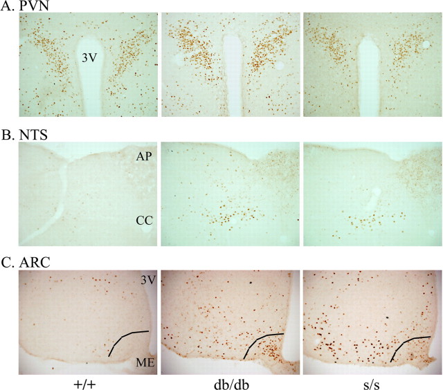
Figure 1.
Regulation of CNS CFLIR by altered LRb action. Immunohistochemical detection of CFLIR in fed +/+, db/db, and s/s mice. A, Representative sections of the PVN at bregma level −1.8 mm. B, Representative section of the NTS at bregma level −13.7 mm. C, Representative sections of the ARC at bregma level −3.0 mm, demonstrating increased CFLIR in the ARC of db/db and s/s mice compared with +/+ mice. The ventromedial ARC (right of line) of db/db mice exhibits a dense population of CFLIR-positive neurons that is essentially absent in s/s mice. CFLIR–DAB-positive neurons are visible as brown nuclei. 3V, Third ventricle; AP, area postrema; CC, central canal; ME, median eminence. Photos taken at 10× magnification.

