Figure 4.
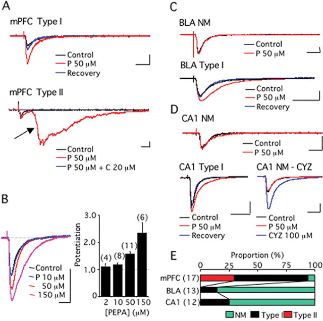
PEPA more potently activates the neural circuit in the mPFC than in the BLA or hippocampal CA1 field. A, mPFC-type I, An example of type I modulation by PEPA of synaptic currents recorded from layer V pyramidal cells in response to electrical stimulation of the mPFC layer II in mouse brain slices. A, mPFC-type II, An example of type II modulation by PEPA of similar synaptic currents. The arrow indicates epileptiform activity. C indicates CNQX. B, Dose-dependent augmentation of synaptic currents by PEPA in cells showing type I modulation. C, BLA-NM, An example of synaptic currents that were not affected by PEPA. NM, no modulation. The synaptic currents were recorded from a BLA pyramidal cell in response to electrical stimulation of the external capsule in mouse brain slices. BLA-type I, An example of type I modulation by PEPA of similar synaptic currents. D, CA1-NM, An example of synaptic currents that were not affected by PEPA. The synaptic currents were recorded from a neuron in the CA1 pyramidal layer in response to electrical stimulation of stratum radiatum in mouse hippocampal slices. CA1-type I, An example of type I modulation by PEPA of similar synaptic currents. NM-CYZ, An example of similar synaptic currents that were not affected by PEPA but potently enhanced by CYZ. Calibrations: 20 ms, 100 pA. E, Summary of the action of PEPA (50 μm) in the mPFC, BLA, and CA1-field (the total number of cells tested is shown in parentheses). The proportion of the cells exhibiting NM, type I, and type II modulations is indicated by color in each bar. Error bars indicate + SEM.

