
Figure 1.
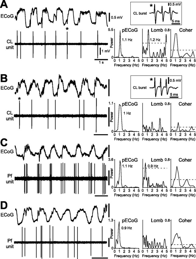
Spike-firing patterns of identified CL and Pf thalamic neurons during cortical SWA. A, Most CL neurons fired bursts of spikes in time with the cortical slow oscillation (∼1 Hz), as shown in the ECoG. Bursts were fired on most oscillatory cycles and were characterized by two to five spikes fired in rapid succession, with progressive attenuation of spike amplitude (a typical burst is denoted by an asterisk and shown in the boxed inset). Comparison of the power spectrum of the ECoG (pECoG) with the Lomb periodogram (Lomb) shows a similar frequency of rhythmic activity in the cortex and the CL neuron spike train. Units of ECoG power are mV2 × 10−2 in all spectra. The dashed line in this and subsequent Lomb periodograms denotes a significance level of p = 0.05. A plot of the coherence between the ECoG and the CL neuron spike train (Coher) showed that these two signals were significantly coherent at ∼1 Hz. The dashed line in the coherence spectrum denotes a significance level of p = 0.05. B, The burst firing of a minority of CL neurons was not rhythmic (peaks in Lomb periodogram did not reach significance level). C, D, Spike-firing patterns of Pf neurons were also heterogeneous during the cortical slow oscillation, with neurons exhibiting either a significant oscillation in firing in time with cortical activity (C) or a nonoscillatory, irregular firing pattern (D). Note that Pf neurons did not usually exhibit the burst firing typical of CL neurons. Calibration in A also applies to B–D.