Figure 3.
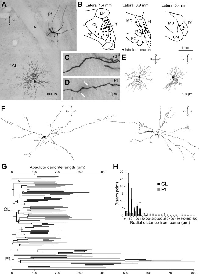
Somatodendritic morphological characteristics of CL and Pf neurons are distinct. A, Montage of light micrographs of a CL neuron and a Pf neuron that were juxtacellularly labeled with Neurobiotin during different electrode penetrations in the same animal. fr, Fascicularis retroflexus. Montage created by merging micrographs from two adjacent sections. B, Locations of all labeled CL and Pf neurons plotted in the sagittal plane. CM, Central medial thalamic nucleus; LP, lateral posterior; MD, mediodorsal; PC, paracentral. C, D, Light micrographs of single dendrites of the neurons shown in A. Filamentous spines (white arrows) are present on the dendrite of the Pf neuron in D but not the CL neuron in C. Scale bar in D also applies to C. E, Digital reconstruction of the entire somatodendritic tree of another labeled CL neuron in the sagittal plane (left) and coronal plane (right). The CL neuron shown is the same as in Figure 1B. F, Digital reconstruction of the entire somatodendritic tree of another Pf neuron in the sagittal plane (left) and coronal plane (right). Note that neurons in E and F are shown at the same scale. G, Dendrograms of a representative CL neuron (top) and a Pf neuron (bottom; same neuron as in F). Note the differences in the number and length of dendritic segments for each neuron type. H, Sholl analysis of dendritic branching as a function of radial distance from soma for two representative CL neurons and five Pf neurons (also see Table 1). Data are means + SEMs for each neuron type. C, Caudal; D, dorsal; R, rostral; V, ventral; M, medial; L, lateral.

