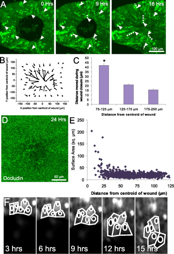
Figure 2.

Closure of lesions in embryonic utricles occurs via movement and cell shape change of cells at the edge of the lesion. A, Frames from a time-lapse recording of a closing of a lesion in the utricle from an E18 transgenic mouse that expresses GFP under control of the β-actin promoter. The GFP is expressed in a mosaic pattern, allowing tracking of individual cell movements (arrows). The trajectory of the cells in the 9 and 18 h images is marked with a dashed line. Cells well outside the lesion moved little during closure of lesions, whereas cells at the leading edge of the lesion moved into the center of the lesion. B, A vector plot showing the distance and direction of movement of 59 cells whose positions were tracked over 15 h of closure of a lesion in a P0 GFP mouse utricle. The cells began at the back of the arrows and moved to the tip of the arrowheads over the 15 h, with each arrow representing the movement of a single cell in the epithelium. The dashed line indicates the initial location of the lesion. Cells at the edge of the lesion moved substantially inward, whereas cells distant from the lesion moved less. C, Histogram of distance moved from 75 cells from each of six E18 GFP mosaic utricles (tracked as in B) plotted based on the original distance from the centroid of the lesion (mean ± SEM). Cells at the edge of the lesion, between 75 and 125 μm of the centroid of the lesion, move significantly farther than cells >125 μm from the lesion (*p < 0.005). D, Fluorescent micrograph showing occludin labeling of the tight junctions between the epithelial cells in an E18 utricle fixed 24 h after lesioning. The occludin staining demarcates the lateral edge of the apical surface of the cells, revealing the expansion of supporting cells at the center of the lesion. E, Comparison of planar surface area versus distance to the center of the lesion for 627 cells in an E18 utricle, showing that cells that moved into center of the lesion site took on greatly expanded apical surface area, whereas more distant cells retained a compact shape. F, Tracings of five cells at the leading edge of the lesion of an E18 GFP mosaic utricle taken every 3 h showing their expansion as the lesion closes.
