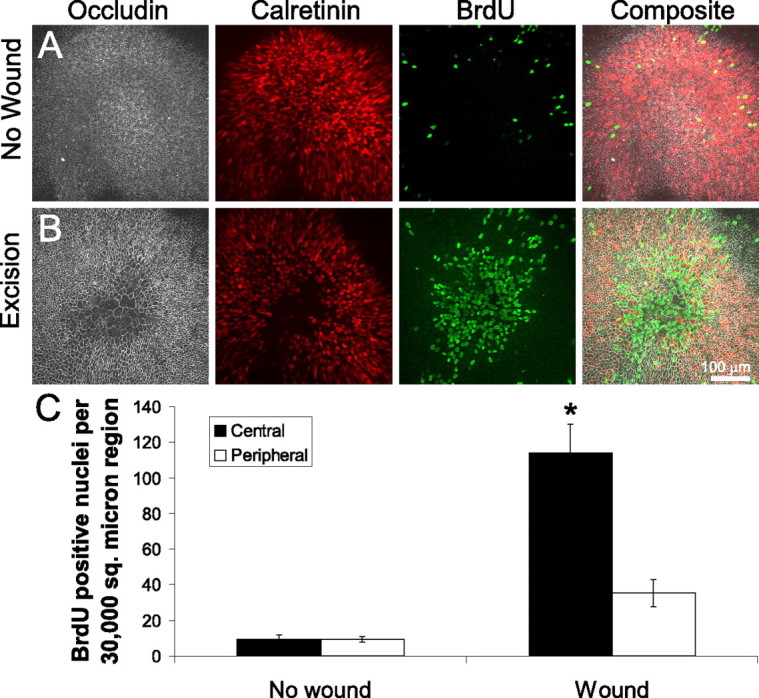
Figure 3.

Cells at the lesion site proliferate after wound closure in embryonic mouse utricles. A, Immunolabeling of an unlesioned E19 mouse utricle cultured for 72 h. Anti-occludin shows the apical surface area of the cells, and anti-calretinin labels the hair cells. Anti-BrdU labels the nuclei of cells that have passed through S-phase during the 72 h of culturing. Few BrdU-positive cells are found within the sensory epithelium. B, Immunolabeling of an E19 mouse utricle cultured for 72 h after excision lesioning. The lesion region in the center of the epithelium shows supporting cells with expanded surface area and extensive BrdU incorporation. C, Quantification of BrdU-positive nuclei in two 30,000 μm2 regions for unlesioned and lesioned utricles: a central circular region over the lesion or center of the unlesioned utricle, and a concentric peripheral region (mean ± SEM). There are few BrdU-positive nuclei in the unlesioned utricles in either the central or peripheral regions, but there is a significant increase in the number of BrdU-positive nuclei in the central region of lesioned utricles (*p < 0.05).
