
Figure 4.
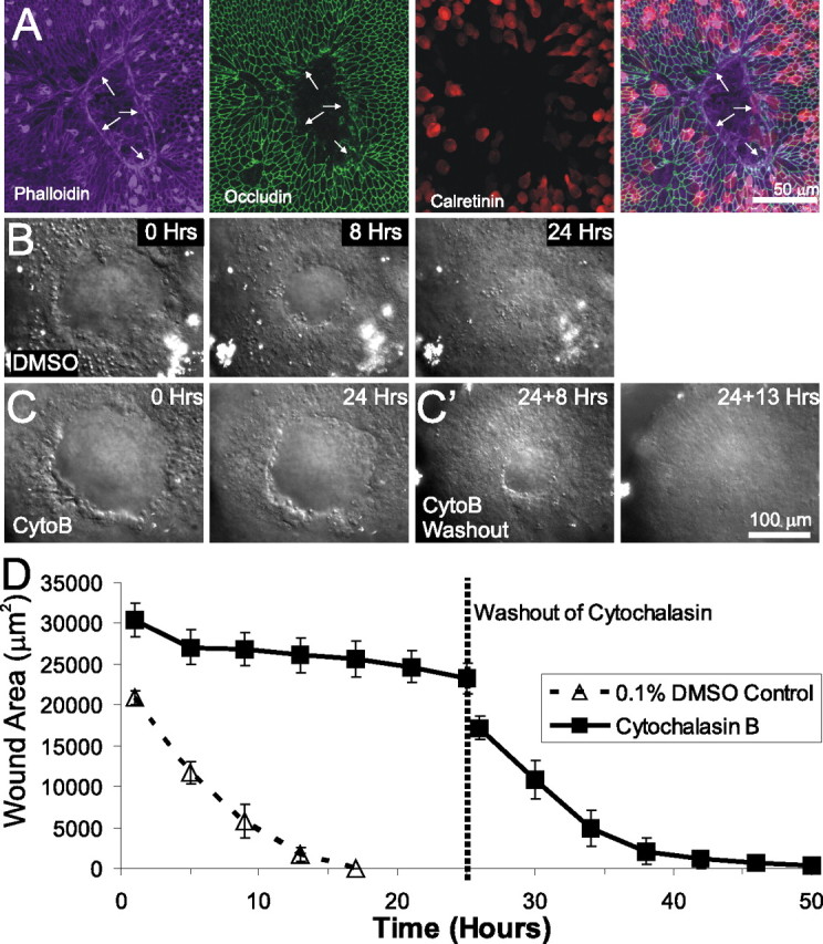
Lesion closure in embryonic utricles occurs via actin purse-string contraction. A, An E18 utricle fixed 6 h after lesioning and triple labeled for phalloidin to label actin, anti-occludin to label tight junctions, and anti-calretinin to label hair cells. An actin purse string runs along the leading edge of the lesion (arrows). B, Individual frames from a time-lapse series showing closure of lesions in a utricle cultured in control media containing 0.1% DMSO. The lesion closed within 24 h. C, Frames from a time-lapse series showing lesion closure in a utricle cultured in media containing 1 μm cytochalasin B (CytoB). After 24 h, the lesion had neither rounded up nor closed. C', After washout of cytochalasin B containing media and replacement with control media, the lesion shown in C contracted and closed 13 h after washout. D, Quantification of lesion size from simultaneous time-lapse recordings of utricles cultured in DMSO control media (dashed line) or cytochalasin B (solid line). The mean ± SEM are plotted every 4 h, showing that lesion closure is inhibited by cytochalasin B but recovers after washout of cytochalasin B.