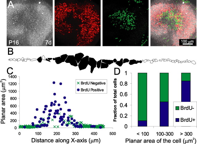
Figure 9.
In mature utricles that have been treated with LPA, proliferation is strongly correlated with whether the cells have an expanded planar area. A, A utricle from a P16 mouse, lesioned and cultured for 7 d in 10 μm LPA, labeled with anti-occludin (white), anti-BrdU (green), and anti-calretinin (red). Many of the cells at the center of the lesion had reentered the cell cycle. B, Representative tracing of cell surface areas based on occludin labeling for a single line of cells from a P16 mouse utricle cultured as in A. Filled cells had a BrdU-positive nucleus, whereas outlined cells did not have a BrdU-positive nucleus. C, Composite plot of cell surface area versus distance along the x-axis for 303 cells from three utricles traced as in B, revealing that BrdU-positive cells were predominantly the large cells covering over the lesion (filled circles represent BrdU-positive cells, and × signs represent BrdU-negative cells). D, A histogram showing the percentage of cells that are BrdU-positive for three bins of cell area. A total of 90% cells with a surface area <100 μm2 were BrdU negative, whereas 85% cells with a surface area >300 μm2 were BrdU positive.

