
Figure 3.
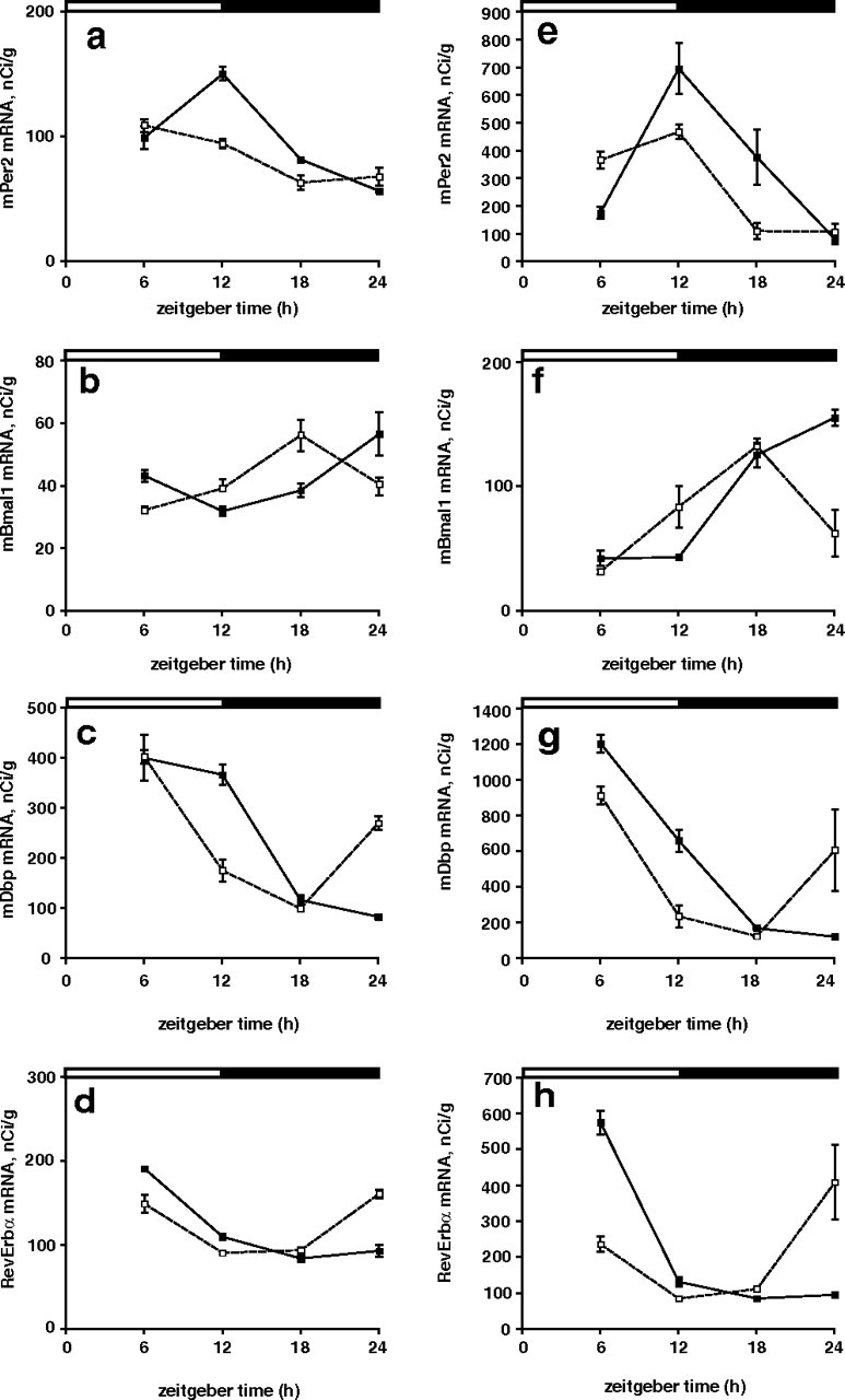
a–h, Rhythms of clock gene expression in the heart (a–d) and liver (e–h) of wild-type and Vipr2−/− mice in LD. Rhythms of Per2 (a, e), Bmal1 (b, f), Dbp (c, g), and RevErbα (d, h) gene expression in wild-type (filled squares) and Vipr2−/− (open squares) mice are shown. Data were obtained by in situ hybridization. The bars at the top indicate the dark period in black and the light period in white. Values represent mean ± SEM.