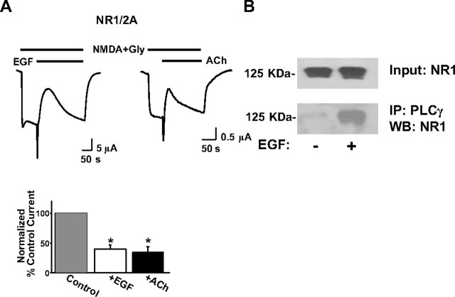
Figure 2.
Stimulation of hormonal receptors that activate PLC transiently inhibits NMDA currents. A, Representative traces of the effect to the NR1/2A current of 100 ng/ml EGF or 5 μm ACh applied as shown. Bars represent mean ± SE percentage current of current before (control current, taken as always 100% before EGF or ACh application in this and subsequent figures when comparison with inhibited states is made) or of maximally inhibited current after EGF or ACh application. Both means representing inhibited current were significantly different from control (*p < 0.001, t test). B, Xenopus oocytes expressing NR1/2A subunits and EGFR were stimulated with EGF (100 ng/ml) as indicated, and membrane preparations were obtained as described in Materials and Methods. Top, Western immunoblot (WB) of a sample of the membrane preparations with an antibody against NR1. The rest of the same membrane preparations were subjected to immunoprecipitation (IP) using an antibody against PLCγ (see Materials and Methods). Bottom, NR1 Western blotting of oocyte membrane immunoprecipitates pulled down with PLCγ antibodies. Blots are representative of two independent experiments with similar results.

