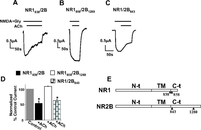
Figure 6.
α-Actinin-binding sites within the C0 domain of NR1 and the distal α-actinin-binding region of the C terminus of NR2B are required for inhibition of NR1/2B currents. A–C, Traces represent recordings of NMDA currents from thapsigargin-treated oocytes expressing wild-type NR1 or NR1 deletion mutants, together with wild-type NR2B or an NR2B mutant as indicated. EGF (100 ng/ml) or ACh (5 μm) were applied as indicated. The constructs have been named after the last residue expressed in the mutant subunit (see Results). D, Bars represent mean ± SE percentage current of current before (control current) or of steady-state current after EGF or ACh application. The means were significantly different only for NR1 or NR2B subunit combinations that contained an α-actinin-binding site (p < 0.01, t test). D, Schematic of NR1 and NR2B subunits with sites of mutants used. α-Actinin binds residues 858–863 of NR1 (see also Fig. 5) and the distal C terminus of NR2B. Gly, Glycine; TM, transmembrane domain.

