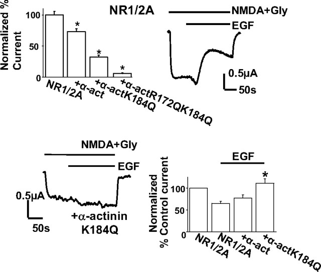
Figure 7.
Mutation of the PIP2-binding residues of α-actinin affects NMDA currents in oocytes. Top left, Bars representing mean ± SE percentage steady-state normalized NMDA current recorded from oocytes expressing NR1/2A together with endogenous α-actinin (control), overexpressed wild-type α-actinin 2, single-point mutant α-actinin 2 K184Q, or double-point mutant α-actinin 2 R172Q–K184Q. The means were significantly different from control (p < 0.001 for all comparisons, t tests). Top right, Representative NMDA current trace of a control oocyte expressing NR1/2A in response to EGF (1 ng/ml) applied as indicated. Bottom left, Representative NMDA current trace of an oocyte expressing NR1/2A and single-point mutant α-actinin 2 K184Q in response to 1 ng/ml EGF. Bottom right, Bars represent mean ± SE percentage current of current before (control current) or of steady-state current after EGF application from oocytes expressing NR1/2A, and NR1/2A together with wild-type α-actinin 2 or single-point mutant α-actinin 2 K184Q. *p < 0.001, statistically significant difference between the NR1/2A group and the K184Q group (t test).

