
Figure 6.
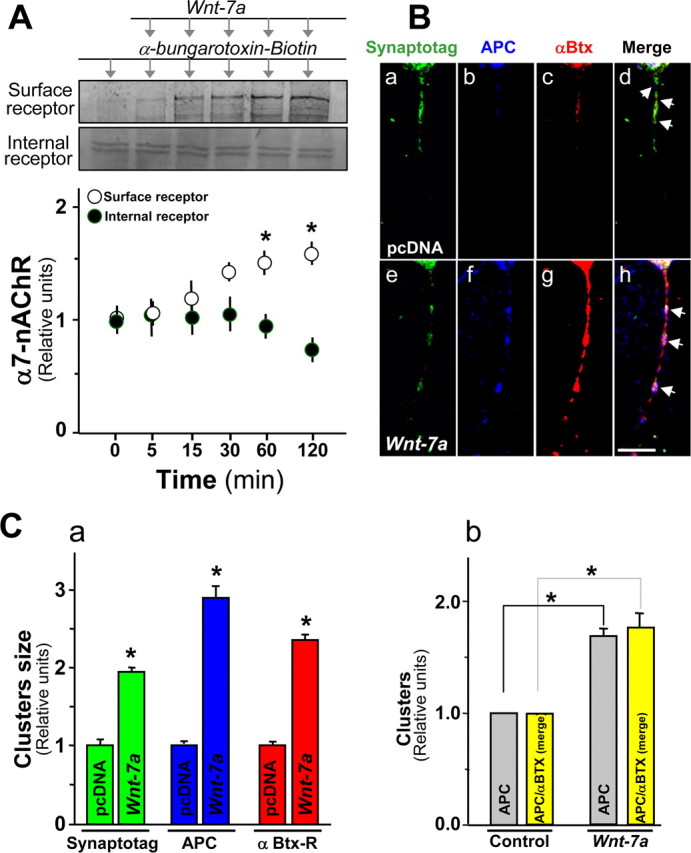
APC and α7-nAChRs are coclustered in presynaptic regions in hippocampal neurons exposed to Wnt-7a. A, Effects on surface α7-nAChR levels assessed using biotinylated-α-Btx pull down and Western analysis in hippocampal neurons subjected to Wnt-7a treatments for different times show increases under conditions in which the fraction of α7-nAChRs in the intracellular pool also assessed by immunoblot decreases (n = 4). B, Rhodamine-labeled α-Btx fluorescence (Bc, Bg) or immunofluorescence for APC (Bb, Bf) or synaptotagmin (Synaptotag; Ba, Be) in hippocampal neurons treated with control medium (pcDNA; Ba–Bd) or Wnt-7a (Be–Bh) for 1 h indicates that Wnt-7a induces clustering of synaptotagmin (Be), APC (Bf), and α7-nAChR (Bg). Bh, Arrows, Wnt-7a elevates coclustering of APC and α7-nAChR in presynaptic regions. Scale bar, 5 μm. C, Quantification of size for synaptotagmin (green bars), APC (blue bars), or α7-nAChR (identified by α-Btx-R staining; red bars) clusters contained in neurites of hippocampal neurons (Ca) and quantification of cluster numbers for APC and for APC contained in α-Btx clusters (Cb). Data are the mean ± SEM of five independent experiments. One hundred clusters per treatment per each independent experiment were evaluated using LSM 5 Image Browser.