
Figure 8.
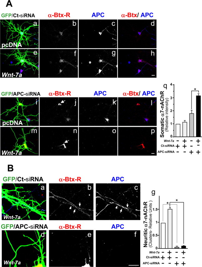
Wnt-7a treatment increases nAChR α7 subunit levels in an APC-independent manner and alters the neuritic localization of α7-nAChRs in an APC-dependent manner. A, Mouse hippocampal neurons cotransfected at 10 d in vitro with green fluorescent protein and control siRNA (GFP/Ct-siRNA; Aa–Ah) or with GFP and anti-APC siRNA (GFP/APC-siRNA; Ai–Ap) were treated in the presence of control media (pcDNA; Aa–Ad, Ai–Al) or Wnt-7a (Ae–Ah, Am–Ap) for 24 h and were stained with rhodamine-labeled α-Btx (α-Btx-R; Ab, Af, Aj, An) or anti-APC (Ac, Ag, Ak, Ao). Arrows show regions of α-Btx-R stain. GFP images were overexposed to visualize neurites. Scale bar, 10 μm. Aq, Somatic levels of α7-nAChR staining. B, Zoom images of Wnt-7a-treated (24 h) GFP/Ct-siRNA (Ba–Bc) or GFP/APC-siRNA (Bd–Bf) mouse hippocampal neurons stained with α-Btx-R (Bb, Be) or anti-APC (Bc, Bf) as well as a quantitation of neuritic α7-nAChR clusters (Bg). Arrows show clusters of α7-nAChR and APC in control transfected neurons. Aq, Neurons transfected with APC siRNA show an increase in the soma of α-Btx-R stain in the presence of control media relative to levels in neurons transfected with control siRNA, and Wnt-7a treatment induces an additional increase in somal α-Btx-R staining in neurons transfected with APC siRNA but not in neurons transfected with control siRNA. Bg, In neurons transfected with inactive siRNA and exposed to Wnt-7a, an increase in the neuritic localization of α7-nAChR is observed relative to similarly transfected neurons in control media, but neuritic α7-nAChRs are very low and insensitive to Wnt treatment in APC-deficient cells. Data are the mean ± SEM of three independent experiments performed in triplicate, expressed as fold increase over control cells.