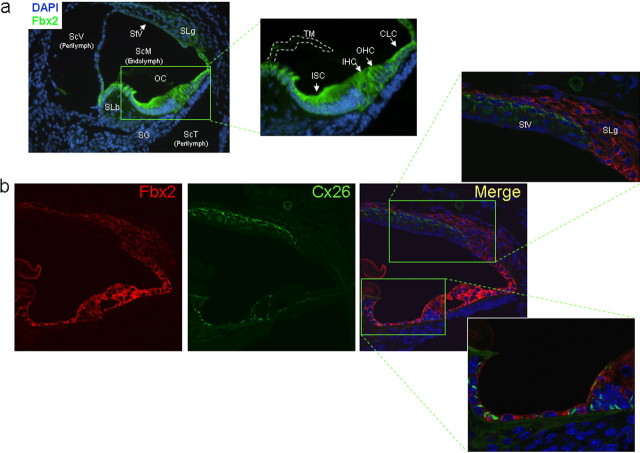
Figure 2.
Fbx2 is highly expressed in the organ of Corti. a, IF analysis for Fbx2 expression (green) in postnatal day 9 mouse cochlea using fluorescence microscopy shows localization to cells of the organ of Corti (OC), including inner sulcus cells (ISC), inner hair cells (IHC), outer hair cells (OHC), and Claudius' cells (CLC), as well as to the spiral ligament (SLg). Fbx2 expression is much less robust in the stria vascularis (StV) and spiral limbus (SLb). Also shown are the three distinct compartments of the cochlea: the scala vestibuli (ScV) and scala tympani (ScT), which contain low-potassium perilymph, and the scala media (ScM), which contains high-potassium endolymph. Spiral ganglion (SG) nerve fibers, which innervate the hair cells, and the tectorial membrane (TM) are labeled. DAPI, Nuclear stain. b, Coimmunofluorescence analysis for Fbx2 expression (red) and Cx26 (green) in postnatal day 14 mouse cochlea using confocal microscopy. Insets, Higher-power confocal imaging shows focal, gap junction staining for Cx26 and cytoplasmic staining for Fbx2 in the organ of Corti support cells. Connexin 26 and Fbx2 do not show similar expression patterns in the StV or SLg. TO-PRO (blue), Nuclear stain.

