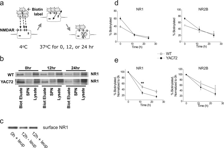
Figure 3.
More rapid degradation of surface NR1/NR2B in cultured striatal MSNs from YAC72 versus wt mice. a, Scheme of biotinylation experiment to analyze receptor degradation. Striatal neurons at 9–10 d.i.v. were incubated with NHS-SS-Biotin for 12 min at 4°C to label surface proteins, washed extensively, and then replaced in their original culture media at 37°C, 5% CO2. Neurons were then harvested at 0, 12, or 24 h after biotinylation, and total soluble lysate was incubated with NeutrAvidin beads to precipitate biotin-tagged proteins, which were then resolved by SDS-PAGE and subjected to immunoblotting. b, Representative immunoblot showing degradation of biotin-tagged NR1. Biot Eluate, Biotinylation eluate; SPN, supernatant. c, Degradation of surface NR1 occurs via the lysosomal pathway. Surface NR1 levels in 9–10 d.i.v. striatal neurons were assessed at 0 or 12 h after biotinylation, in the presence (+leup) or absence (−leup) of 100 μg/ml leupeptin (lysosomal inhibitor). Blot shown is representative of three independent experiments from different batches of wt or YAC72 MSNs. d, Plot of degradation of biotin-tagged NR1 (left) or NR2B (right) over time. Values represent mean ± SEM from n = 5–12 (NR1) or 2–3 (NR2B) independent experiments with different batches of cultured neurons. e, Plot of degradation of biotin-tagged NR1 (left) or NR2B (right) with values of the 0 h time point in each genotype normalized to 100%. Values represent means normalized to 100% at 0 h ± SEM. Significantly different compared with wt, **p < 0.01 by unpaired t test.

