Figure 3.
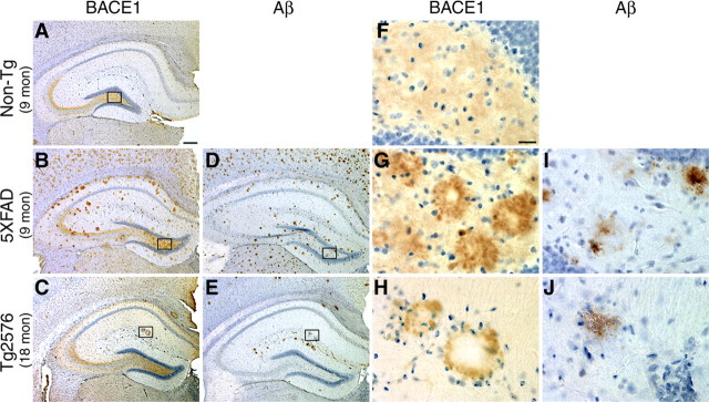
BACE-Cat1 reveals plaque-like staining in APP transgenic brains by immunohistochemistry. A–J, Coronal brain sections from 5XFAD and Tg2576 mice were immunostained with BACE-Cat1 antibody (A–C, F–H) or anti-total Aβ-amyloid antibody 4G8 (D, E, I, J) and counterstained with hematoxylin. Only images of the hippocampus are shown. A, F, Nontransgenic (Non-Tg) brain section from 9-month-old mouse stained with BACE-Cat1 shows the normal BACE1 immunoreactivity pattern with strongest labeling in the hippocampal mossy fiber pathway. B, G, 5XFAD brain section from 9-month-old mouse stained with BACE-Cat1 displays plaque-like BACE1 immunoreactivity. Note that BACE1-positive deposits typically have unstained cores. D, I, Adjacent 5XFAD brain section stained with 4G8 antibody reveals amyloid plaques that have a similar spatial distribution as BACE-Cat1-positive deposits. C, H, Tg2576 brain section from 18-month-old mouse stained with BACE-Cat1 also shows a plaque-like pattern of BACE1-positive deposits, although the number of deposits is less than in 5XFAD mice. E, J, Adjacent Tg2576 brain section immunostained with 4G8 antibody shows a similar distribution of amyloid plaques as that of BACE1-positive deposits. F–J, Higher magnification images of boxed areas in A–E. BACE1 immunoreactivity typically forms a ring-like structure with a clear center in both 5XFAD and Tg2576 transgenic mice (G, H), whereas amyloid plaques have a more uniformly stained core (I, J). Scale bars: A–E, 200 μm; F–J, 20 μm.

