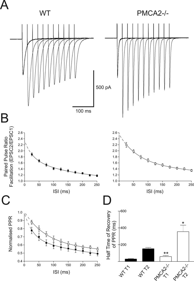
Figure 1.
PF-evoked EPSCs in PNs from PMCA2−/− mice exhibited enhanced PPF that recovered more slowly than WT PPF. A, PPF of the PF input to PNs from WT (left) and PMCA2−/− (right) mice, at varying ISIs. Traces represent responses averaged over 17 cells. B shows the average PPR as a function of ISI for all cells, WT (filled symbols and lines) and PMCA2−/− (open symbols), where dotted lines show extrapolated values determined from the exponential fits. C shows recovery of amplitude normalized PPR for WT and PMCA2−/− (filled and open symbols and lines, respectively). Recovery of PPR fitted with a double exponential in all cases. The first half-time for PPR decay, T1, increased from 21 to 51 ms and the second, slower half-time for PPR decay, T2, increased from 286 to 650 ms. D shows means from individual recovery phases of PPR (T1 and T2) for WT (filled bars) and PMCA2−/− (open bars). *p < 0.05; **p < 0.01. All values are mean ± SEM.

