
Figure 6.
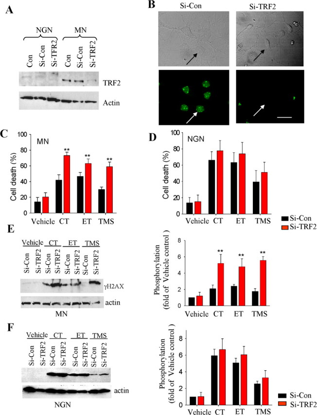
RNA interference-mediated depletion of TRF2 renders mature neurons vulnerable to telomere and DNA damage. NGNs and MNs were infected with a lentiviral vector containing shRNA directed against TRF2 mRNA (Si-TRF2) or a control lentiviral vector with a nonsense shRNA sequence (Si-Con); additional cells were not exposed to virus (Con). A, At 48 h after infection, cell lysates were subjected to immunoblots using antibodies against TRF2 and β-actin. B, At 48 h after infection, cells were immunostained with TRF2 antibodies. Arrows show the same cells in transmitted light and immunofluorescence images. C, D, MNs expressing TRF2 siRNA exhibit increased vulnerability to cell death induced by telomere and DNA-damaging agents, whereas the vulnerability of NGNs is unaffected by TRF2 siRNA expression. E, F, MNs expressing TRF2 siRNA exhibit increased γ-H2AX levels after exposure to TMS, etoposide (ET), or camptothecin (CT), whereas γ-H2AX levels in NGNs are unaffected by TRF2 siRNA expression. At 48 h after infection, NGNs and MNs were exposed to camptothecin (10 μm), etoposide (10 μm), or TMS (1 μm) for either 24 h (cell death analysis) or 3 h (γ-H2AX analysis). Cell death was quantified by Hoechst dye staining, and phosphorylation of histone H2AX was analyzed by densitometric analysis of γ-H2AX immunoblots. Values are the mean ± SD of three independent experiments. **p < 0.001, paired Student's t test.