Figure 3.

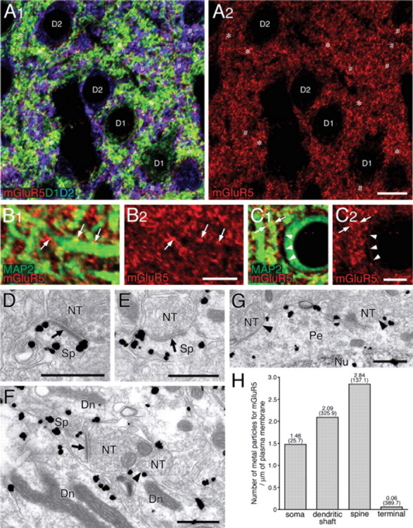
mGluR5 is distributed on somatodendritic elements of D1R-positive and D2R-positive medium spiny neurons. A1, A2, Triple immunofluorescence for mGluR5 (red), D1R (green), and D2R (blue). Note a punctate or reticular pattern of mGluR5 immunolabeling, which is detected on D1R-positive (*) and D2R-positive (#) elements in the neuropil. B1–C2, Double immunofluorescence for mGluR5 (red) and MAP2 (green). In addition to dense mGluR5 labeling in the neuropil, low to moderate labeling can be detected on the surface of shaft dendrites (arrows) and somata (arrowheads) of presumed MS neurons. D–G, Silver-enhanced immunogold showing preferential cell surface distribution of mGluR5 in spines (Sp), dendritic shafts (Dn), and perikarya (Pe) of presumed MS neurons. Arrows and arrowheads indicate asymmetrical and symmetrical synapses, respectively. NT, Nerve terminal; Nu, nucleus. H, The mean number of metal particles for mGluR5 per 1 μm of the cell membrane. From very low density in terminals, mGluR5 is judged to be specific to somatodendritic compartments, similarly to DAGLα. Numbers in parentheses indicate the total length of measured cell membrane. Scale bars: A2, 10 μm; B2, C2, 2 μm; D–G, 500 nm.
