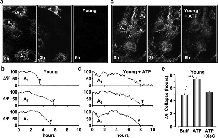
Figure 1.
P2Y-R activation protects young astrocytes from oxidative stress. a, Images of astrocytes cultured from young mice labeled with the mitochondrial potential-sensitive dye TMRE and exposed to oxidative stress for the indicated times. Each image is a maximum intensity projection of a z-stack of six optical sections (1 μm steps). Plated cells were continuously perfused at 37°C with culture medium containing TMRE (200 nm) and imaged with two-photon microscopy (800 nm). b, Line plots of the mean TMRE fluorescence (ΔΨ) for the cells labeled in a. Fluorescent units are scaled to 100% at 0 h. The times when the TMRE fluorescence collapses to 10% of the initial values are indicated by black arrowheads. c, Images of young astrocytes pre-exposed to extracellular ATP (10 μm) for 10 min before adding t-BuOOH (100 μm) to the perfusate (0 h). d, Line plots of the TMRE fluorescence for the cells indicated in c. e, Histograms of the mean times of ΔΨ collapse for young astrocytes exposed to buffer only (Buff), ATP, or ATP plus Xestospongin C. ***p < 0.001.

