
Figure 6.
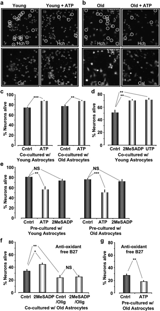
Stimulation of P2Y-Rs in astrocytes enhances their ability to protect neurons from oxidative stress. a, b, Images of cortical neurons cocultured with young (a) and old (b) astrocytes acquired 4 h after incubation with t-BuOOH (100 μm). Cocultures pretreated with ATP (2 μm; 10 min) are presented in the right panels. The top images show Hoechst (Hch) stained nuclei. The bottom images show the same field of neurons stained with calcein (Cal), which only stains the cytoplasm of cells that are alive. Hoechst-labeled nuclei that are calcein negative are circled and counted as cell deaths. c, Histogram plots of the percentage of neurons alive when cocultured with young or old astrocytes. d, Histogram plots of the percentage of neurons alive pretreated with either 2-MeSADP (2 μm) or UTP (50 μm) for 10 min. e, Histogram plots of the percentage of neurons alive that were precultured with astrocytes but were separated before pretreating (10 min) the neurons with either ATP (2 μm ATP; 10 min) or 2-MeSADP (2 μm) and subsequently exposed to oxidative stress (100 μm; t-BuOOH; 4.5 h). f, Histogram plots of percentage of neurons alive cocultured with old astrocytes using anti-oxidant free B-27 supplement for 3 h. g, Same experiment as in e except using anti-oxidant free B-27 supplement. ***p < 0.001; **p < 0.01; *p < 0.05.