
Figure 4.
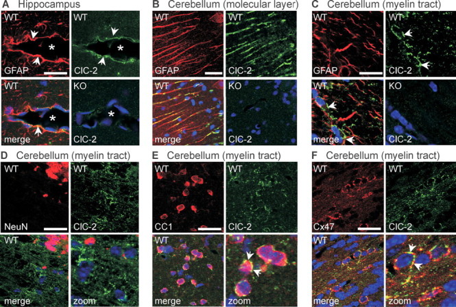
Subcellular localization of ClC-2 in brain. Confocal images of brain sections of 5-week-old WT and ClC-2 KO mice double stained for ClC-2 (green) and marker proteins (red) including GFAP (A–C), NeuN (D), CC1 (E), and Cx47 (F). A, In the hippocampus, ClC-2 colocalized with GFAP in astrocytic endfeet (arrows) surrounding blood vessels (asterisks). B, C, In the cerebellum, ClC-2 was found in GFAP-positive Bergman glia of the molecular layer (B) and in myelinated fiber tracts (C). C, D, In the latter region, ClC-2 did not significantly overlap with either GFAP (C) or NeuN (D). E, Cells with numerous ClC-2 puncta around their cell bodies were identified as oligodendrocytes by staining for CC1. F, Many of these ClC-2-positive puncta also stained for Cx47. Scale bars, 20 μm. Blue indicates TOTO staining of nuclei.