
Figure 2.
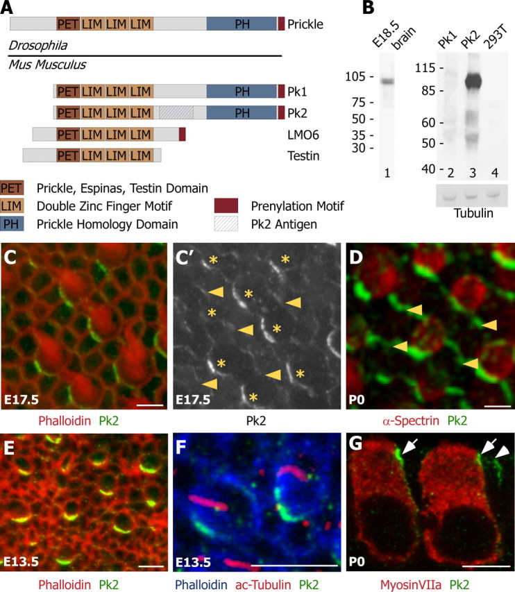
Pk2 is localized asymmetrically at cell boundaries throughout hair cell differentiation and polarization. A, Four Prickle-like proteins can be identified in mouse on the basis of the presence of a single PET and three LIM protein domains. Two of these, Pk1 and Pk2, also contain a Prickle homology domain and are the most similar to Drosophila Prickle. A region unique to Pk2 (hatched box) was used to generate Pk2-specific antiserum. B, On Western blots, the Pk2 antiserum recognizes a single band from E18.5 brain extract (lane 1) and HEK 293T cells expressing Pk2 (lane 3), but not Pk1 (lane 2) or nontransfected HEK 293T cell lysates (lane 4). Immunoblotting with a tubulin antibody was included as a loading control. C, In developing E17.5 utricles, Pk2 (green) is enriched at one edge of hair cells, labeled with phalloidin (red). C′, In the utricle, Pk2 is present at lower levels at boundaries between adjacent support cells than at boundaries between hair cells and support cells. Shown is a gray scale image of Pk2 labeling from C, with arrowheads indicating boundaries between adjacent supporting cells and asterisks marking the positions of hair cells. D, In the saccule, Pk2 labeling (green) is more prominent at boundaries between support cells (arrowheads) than in the utricle. Hair cells are labeled with α-spectrin (red). E, At E13.5, Pk2 (green) is enriched at one edge of hair cells that lack a stereocilia bundle but can be identified by their rounded shape and mosaic distribution via phalloidin stain (red). F, Pk2 accumulation (green) at E13.5 precedes the asymmetric localization of the kinocilium (red) to one edge of the cell (phalloidin; blue). G, In sections, Pk2 enrichment (green) is seen near the apical surface of the tissue at boundaries between supporting cells (arrowhead) and adjacent to hair cells (arrows), identified by the hair cell marker MyosinVIIa (red). Scale bars, 5 μm.