
Figure 3.
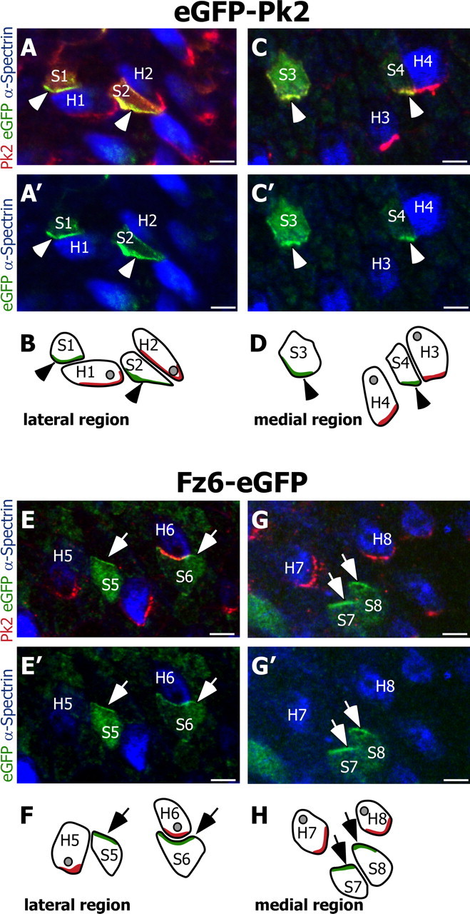
Pk2 and Fz6 are redistributed to opposite sides of utricular hair cells and support cells. A, Two support cells located within the lateral domain of the utricle (labeled S1 and S2) electroporated with eGFP-Pk2 (green) distribute eGFP-tagged Pk2 along their medial edges (arrowheads). Endogenous Pk2 (red) forms crescents along the medial cell boundary of two hair cells [identified by using α-spectrin (blue); labeled H1 and H2] and does not overlap with the distribution of eGFP-Pk2 in S1 or S2. The Pk2 antibody also labels exogenous eGFP-Pk2 in the support cells. Electroporated cells are located in the lateral domain of the utricle. B, Diagram illustrating the relative positions of support cells and hair cells from A and A′. In this and all subsequent diagrams the subcellular localizations of eGFP-tagged protein and endogenous Pk2 are indicated by green and red shading, respectively. The position of the hair cell kinocilia, based on α-spectrin labeling, is illustrated as a gray spot. C, C′, D, Support cells located on the opposite side of the line of reversal and within the medial region of the utricle (labeled S3 and S4) also distribute eGFP-tagged Pk2 (arrowheads) along their medial edges, similar to endogenous Pk2 in hair cells (labeled H3 and H4). E, E′, F, In contrast, support cells electroporated with Fz6-eGFP (labeled S5 and S6) redistribute Fz6-eGFP protein (green) to their lateral edge (arrows). In these cells Fz6-eGFP is enriched opposite of the endogenous Pk2 crescents (red) present in adjacent hair cells (H5 and H6). In E and F the electroporated cells are located in the lateral domain of the utricle. G, G′, H, Support cells located on the opposite side of the line of reversal and within the medial region of the utricle (labeled S7 and S8) also redistribute Fz6-eGFP to their lateral edge (arrows). This distribution is opposite from that of the endogenous Pk2 (red) in hair cells (labeled H7 and H8). For each utricle, the medial and lateral regions are identified by the position of the line of reversal, and images are oriented with the lateral edge of hair cells on top. Scale bars, 5 μm.