
Figure 1.
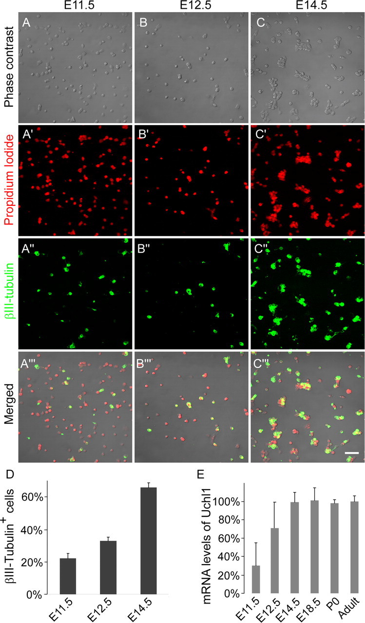
Cultured DRG cells from all rostrocaudal levels at E11.5, E12.5, and E14.5 stained with the general nuclear marker propidium iodide (A′–C′, red) and for the neuronal marker βIII-tubulin (A″–C″, green; A‴–C‴, merged), showing the gradual increase in neuronal differentiation. Scale bar, 50 μm. D, Quantification of A–C (n = 3). E, mRNA levels of the neuronal marker UCHL1, normalized against the adult.