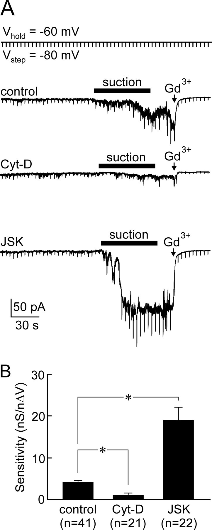
Figure 3.

Role of F-actin in mechanotransduction. A, Whole-cell current recordings (VH = −60 mV) from isolated MNCs exposed to Cyt-D (middle), vehicle (control, top), or JSK (bottom). Vertical deflections are current responses to hyperpolarizing steps (−20 mV) applied every 5 s to monitor G. In each case, negative pressure (∼−100 mmHg) was applied to the patch pipette (bar) to induce a decrease in cell volume of ∼10%. Note that Gd3+ (250 μm) was applied near the end of each recording to confirm that the current induced was caused by the activation of SIC channels. B, Bar graph showing mean (±SEM) values of transducer mechanosensitivity measured in each group (*p < 0.05; ANOVA).
