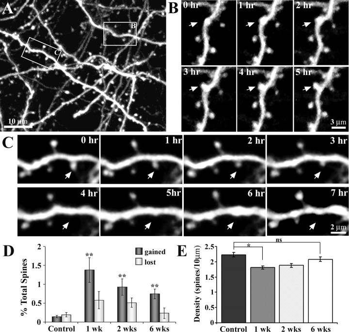
Figure 4.
Time-lapse imaging of apical dendrites reveals heightened levels of spine formation in peri-infarct cortex. A, Maximal intensity z projection of 30 planar images (taken 1 μm apart, at 0 h) depicting the Golgi-like detail of YFP-labeled dendrites. In this field of view, a dendritic spine formed (box labeled B) and another retracted (box labeled C) over the imaging period. B, C, Time-lapse images (taken 1 h apart) showing the formation and retraction, respectively, of a dendritic spine. D, Relative to controls, the rate of spine formation was significantly increased in peri-infarct cortex at 1, 2, and 6 weeks after stroke. Spine elimination, although elevated, was not significantly different from control levels. E, Relative to controls, spine density was significantly reduced at 1 and 2 weeks, but appeared to recover by 6 weeks. *p < 0.05; **p < 0.005.

